Dysplastic Nevus (Atypical Mole)
Understanding atypical moles and their risks.
Introduction
Dysplastic nevi, commonly known as atypical moles, are unusual skin lesions that differ from normal moles in several ways. They can vary in color, size, and shape, and their irregular appearance often raises concerns about the potential for skin cancer. Understanding the nature of dysplastic nevi is crucial for effective monitoring and management, as they can sometimes be associated with an increased risk of melanoma.
Moles, or nevi, are clusters of pigmented cells that commonly appear during childhood and adolescence. While most people have moles, not all moles are considered atypical. Dysplastic nevi are distinguished by their atypical features, which necessitate greater awareness and potential medical attention. This guide aims to provide comprehensive information on dysplastic nevi, including their characteristics, causes, diagnosis, and management.
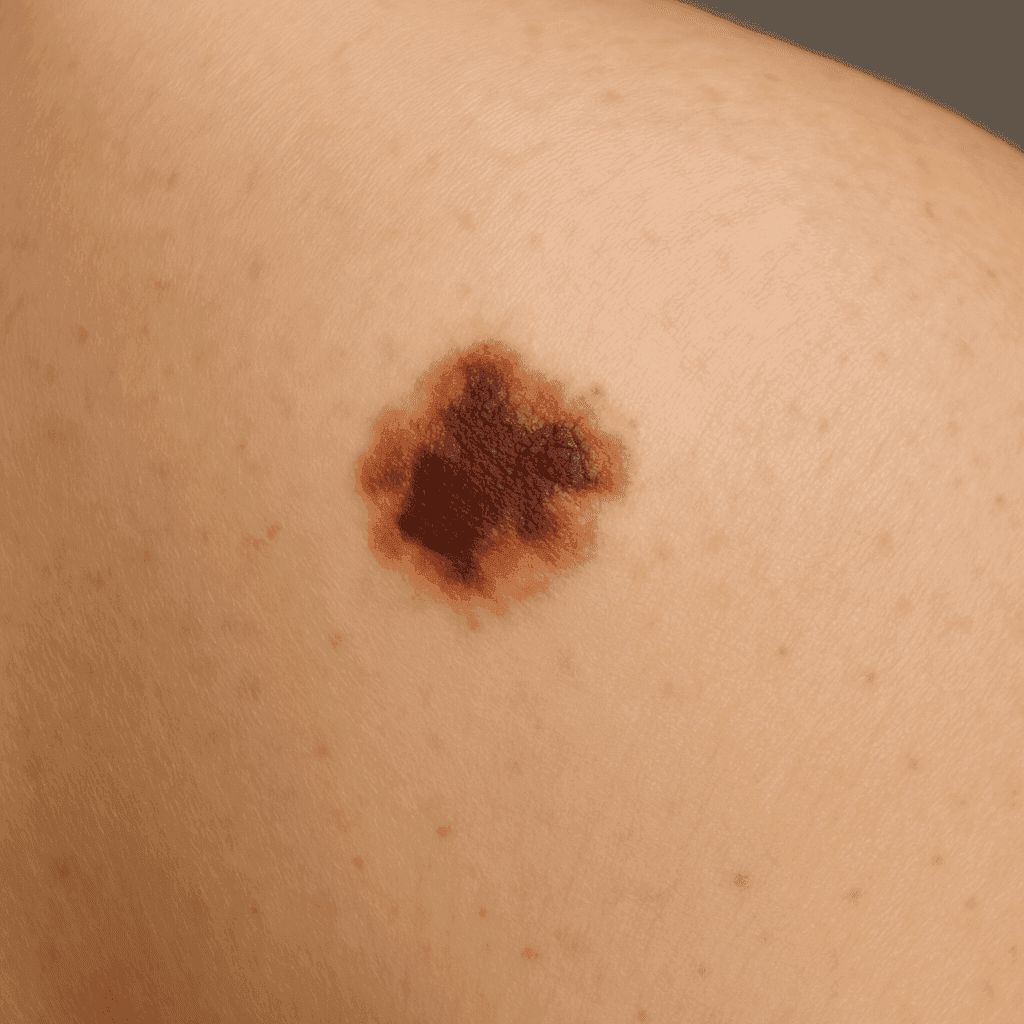
Characteristics of Dysplastic Nevus
Appearance and Features
Dysplastic nevi often have a distinct and irregular appearance compared to normal moles. They may be larger than 5 millimeters in diameter and exhibit a mix of colors, such as various shades of brown, black, and sometimes pink. The borders of these moles can be notched, blurred, or irregular, and their surfaces might be smooth, scaly, or rough.
Differences Between Normal Moles and Atypical Moles
While normal moles are generally uniform in color and have smooth, well-defined borders, atypical moles deviate from these characteristics. They can exhibit asymmetry, where one half of the mole looks different from the other, and this irregularity might suggest a need for further examination. It’s important to note that while dysplastic nevi can resemble melanoma, they are not cancerous, although they may indicate a higher risk for the disease.
Causes and Risk Factors
Genetic Predisposition
Genetics play a significant role in the development of dysplastic nevi. Individuals with a family history of atypical moles or melanoma are more likely to develop these types of moles. Certain genetic mutations have been linked to an increased likelihood of forming dysplastic nevi, making genetic predisposition a key factor.
Environmental Factors
Aside from genetics, environmental influences such as excessive sun exposure can contribute to the formation of atypical moles. Ultraviolet (UV) radiation from the sun or tanning beds can damage skin cells, potentially leading to the development of dysplastic nevi. People with lighter skin, who are more sensitive to UV exposure, are at greater risk.
Dysplastic Nevus and Melanoma Risk
Understanding the Connection
The relationship between dysplastic nevi and melanoma is complex. While dysplastic nevi themselves are not cancerous, their presence can indicate a higher risk of developing melanoma, a serious form of skin cancer. This risk is especially notable in individuals with a high number of atypical moles.
Statistics and Studies
Research has shown that individuals with multiple dysplastic nevi have a significantly higher risk of melanoma compared to those with fewer or no atypical moles. It is estimated that people with dysplastic nevi are at a 10-fold increased risk of melanoma. Regular monitoring and early detection are essential in managing this risk effectively.
Diagnosis of Dysplastic Nevus
Physical Examination
A thorough physical examination by a dermatologist is the first step in diagnosing dysplastic nevi. Dermatologists assess the size, color, and shape of moles, looking for any irregularities that may suggest atypical characteristics.
Dermatoscopy and Imaging Techniques
Dermatoscopy, a non-invasive imaging technique, allows dermatologists to examine moles with greater precision. This tool provides a magnified view of the skin, highlighting patterns and structures not visible to the naked eye. Dermatoscopy can aid in distinguishing dysplastic nevi from melanoma.
Biopsy Procedures
In cases where a mole appears suspicious, a biopsy may be performed. This involves removing a sample of the mole for laboratory analysis to determine whether it is benign or malignant. Biopsies are a crucial step in confirming a diagnosis and guiding treatment decisions.
Treatment Options
Monitoring and Regular Check-ups
For many individuals with dysplastic nevi, regular monitoring and check-ups with a dermatologist are key components of management. This involves keeping track of any changes in the moles over time and scheduling routine skin examinations.
Surgical Removal
If a mole shows signs of change or poses a risk, surgical removal may be recommended. This procedure involves excising the mole and surrounding tissue to ensure complete removal. It is typically performed under local anesthesia and is considered a straightforward and effective treatment.
Non-surgical Treatments
While surgical removal is the primary treatment for dysplastic nevi, preventive measures and regular monitoring remain critical. Non-surgical approaches focus on reducing future risks and involve lifestyle adjustments such as avoiding excessive sun exposure and practicing good skin care.
Prevention and Monitoring
Self-Examination Techniques
Regular self-examinations are an important practice for individuals with dysplastic nevi. This involves checking the skin for new moles or changes in existing ones using the ABCDE rule (Asymmetry, Border, Color, Diameter, Evolution) as a guide.
When to See a Dermatologist
It is recommended to visit a dermatologist at least once a year for a professional skin examination. If you notice any changes in your moles, such as an increase in size, change in color, or irregular borders, seek medical advice promptly.
Lifestyle and Protective Measures
Preventive measures include using broad-spectrum sunscreen with a high SPF, wearing protective clothing, and avoiding peak sun exposure hours. These steps can help reduce the risk of developing new dysplastic nevi and minimize the progression of existing ones.
Dysplastic Nevus Syndrome
Overview of the Condition
Dysplastic nevus syndrome, also known as familial atypical multiple mole and melanoma (FAMMM) syndrome, is a genetic condition characterized by the presence of multiple atypical moles and an increased risk of melanoma. Individuals with this syndrome often have a family history of melanoma and dysplastic nevi.
Management Strategies
Management of dysplastic nevus syndrome involves regular skin examinations, both self-conducted and by healthcare professionals. Genetic counseling may be recommended for families affected by this condition to assess and manage the potential risks. Early detection and intervention are critical in reducing the risk of melanoma.
Frequently Asked Questions
What is the difference between a dysplastic nevus and melanoma?
Dysplastic nevi are atypical moles that may resemble melanoma but are not cancerous. However, they can increase the risk of developing melanoma.
Can dysplastic nevi be prevented?
While genetic factors cannot be changed, reducing sun exposure and using sunscreen can help prevent the development of new atypical moles.
How often should I have my atypical moles checked by a dermatologist?
It is recommended to have regular check-ups at least once a year, or more frequently if advised by your dermatologist.
Is it necessary to remove all dysplastic nevi?
Not all dysplastic nevi need to be removed. Removal is typically considered if there are changes in size, shape, or color, or if recommended by a healthcare professional.
Are there any non-surgical treatments for dysplastic nevi?
Currently, surgical removal is the primary treatment for atypical moles. Other treatments are mainly focused on monitoring and prevention.
What should I do if I notice changes in my moles?
If you observe any changes in your moles, such as asymmetry, border irregularity, color changes, or size increase, consult a dermatologist promptly.